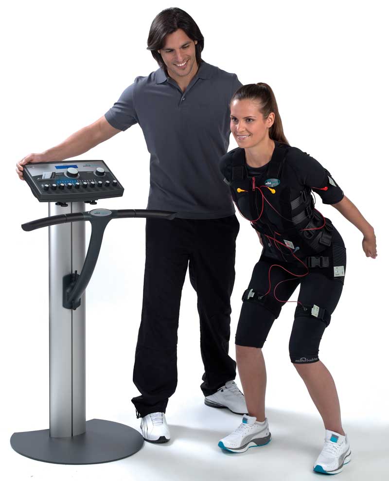
Electromyostimulation (EMS) has enjoyed ever greater popularity in recent years. Short training sessions are supposed to be sufficient to achieve the same effects as conventional strength training with dumbbells – according to advertising promises. This is an appealing idea and fits into the usually tight schedule of modern everyday life. The application of current via the skin is anything but new, however. As long ago as 46 AD, the Roman physician Seribonius Largus used the current generated by electric rays to treat his patients’ headaches and gout (Finger & Piccolino, 2011). Testing the electrical activation of muscles in athletes began in Russia in the 1960s (Ward & Shkuratova, 2002). Considerable time has passed since these early trials and the devices have been subject to continuous further development. This applies in particular to the electrode material used and the possibility of applying current to several muscle groups at the same time by means of special clothing and devices. This has significantly improved the practicability of the application and undoubtedly contributed to the success achieved by EMS in recent years.
Sceptics, however, see in EMS Training an unnatural and therefore potentially dangerous form of training. Statements such as “this simply cannot be healthy” are often heard in this context. Doubts have also been frequently expressed that the contractions generated are actually capable of producing biopositive adaptation phenomena. The aim of this article is therefore to gather information on the benefits and risks of EMS Training.
Background
The electrical stimulation of the muscles leads to an involuntary contraction of the activated muscle fibres. Luigi Galvani recognized this connection in his early experiments on frogs’ legs in the 18th century. He used, among other things, the electricity of thunderstorms to bring about contractions in the frogs’ muscles (Bresadola, 1998). He connected the nerve fibres of the frogs' legs to a kind of lightning rod (see Fig. 1). Thankfully, the current form of EMS Training is no longer reliant on electric arcs between clouds and earth whose electrical energy constitutes several billion joules. The devices used for EMS Training work with energies so low that they are generally harmless to the human body. Exceptions to this are certain conditions where EMS Training should be avoided because of the lack of available study results. Included among these is the use of a heart pacemaker or a pregnancy, for example.
EMS-induced muscle damage
Reports often appear in the press of massive muscle injuries that were triggered by EMS Training in supposedly healthy individuals and which had to be treated in hospital in order to prevent life-threatening consequential damage to internal organs. Acute renal failure in particular is a concern in this regard, as it can be triggered by the inundation of muscle breakdown products. In 2015, for example, Spiegel Online reported on a 48-year-old woman who had complained about circulatory problems, heart palpitations and chest pain after an EMS Training session (Habich, 2015). According to the information provided by Spiegel Online, the lady was unable to get up for several days and received a number of infusions in hospital to protect her kidneys. Diagnosis: Rhabdomyolysis – the damage-induced breakdown of muscle fibres. How is this risk to be assessed?

Fig.: 1 Setup by Luigi Galvani to study the influence of electricity on the contraction of skeletal muscles.
Muscle damage markers and the crush kidney
The extent of muscular damage is usually estimated indirectly in the clinic by means of so-called muscle damage markers. These markers are enzymes and other proteins which are unable to leave the cell interior of the muscle fibres because of their molecular size. If, however, the muscle cell membrane is damaged, these substances enter the extracellular space and from there, either directly or via the lymph system, into the blood circulation. The enzyme creatine kinase (Creatine Kinase; CK) is deemed to be the most sensitive marker of rhabdomyolysis. Although there are no reliable limit values for the activity of CK in the blood, some authors still attempt to define rhabdomyolysis in terms of the serum activity of CK. Values between 170 IU∙L-1 und 1,700 IU∙L-1 are then described as mild, up to 8,330 IU∙L-1 as moderate and beyond that as pronounced rhabdomyolysis. In the above-mentioned Spiegel Online article, the doctors found a CK value of 26,000 IU∙L-1, which would correspond to pronounced rhabdomyolysis. The risk of renal damage does not depend however on the CK, but on the myoglobin which is also released. This is excreted via the kidneys and has a toxic effect on the renal tissue at high concentrations. Such interrelations are known in emergency medicine and are observed, among other things, when large soft tissue traumas occur in the event of accidents. The clinician speaks of renal damage induced by a so-called crush kidney.
Kemmler et al. (Kemmler, Teschler, Bebenek, & Stengel, 2015) devoted themselves in a recently published study to the health relevance of high muscle damage markers in the blood after excessive whole-body EMS. Three to four days after a 20-minute EMS session (85 Hz; 350 ms; intermittent, bipolar), the researchers found CK values of 28,545 ± 33,611 IU∙L-1 and a 40-fold increase in the myoglobin concentration from 68 ± 44 µg∙L-1 to 2,706 ± 2,194 µg∙L-1, which appeared only two to three days after the stimulation. The observed values in the blood were therefore seven times higher than the toxic limit quoted in the literature for myoglobin of 370 µg∙L-1 (El-Abdellati et al., 2013). Despite this, the working group under Kemmler could not find any signs of acute renal failure in any of the subjects. How can this be explained?
Clarkson et al. (Clarkson, Kearns, Rouzier, Rubin, & Thompson, 2006) made similar observations after 50 eccentric contractions of the elbow flexion musculature in 203 test subjects. Despite massive CK increases of up to 80,550 IU∙L-1 and Mb concentrations of up to 3,200 µg∙L-1, the authors found no significant impairment of renal function. Sinert et al. (Sinert, Kohl, Rainone, & Scalea, 1994) were unable to find any evidence of acute renal failure in the patients they examined with pronounced rhabdomyolysis (40,471 ± 34,295 IU∙L-1). This is in contrast to the 17 - 40 % incidence of acute renal failure associated with rhabdomyolyses which is described in the literature (Akmal, Valdin, McCarron, & Massry, 1981; Ward, 1988). That may indicate that other nephrotoxic factors must be present in order for the feared complication of myoglobin elevation to develop. Even if this data shows that even massive increases in the designated muscle damage markers in the blood may not have any health consequences, it must be taken into account that any pre-existing illnesses of the customers in the EMS fitness studio can significantly reduce the tolerance threshold of the kidney for Mb. Against this background, such high marker elevations are in any case to be avoided for safety reasons.
The “Repeated Bout Effect”
It is noticeable that these high values are usually reached only in the initial training sessions. That is why, in the above-mentioned study by Kemmler et al. (Kemmler et al., 2015), it was possible to show that an EMS Training session produced significantly lower CK values (906 ± 500 IU∙L-1) after a 10-week training phase than were produced in the first session (17,575 ± 14,717 IU∙L-1). This makes a difference of 16,669 IU∙L-1. The myoglobin elevation was also significantly lower at 193 ± 80 µg∙L-1. This effect, which is called the “Repeated Bout Effect”, describes a kind of protective mechanism of the body from further stress and can also be observed in other forms of training. While it has not yet proved possible to clarify the mechanism involved in the Repeated Bout Effect, it is known however that the effect is not limited to trained muscle groups, but is also cross-transferred to non-trained muscle groups (Starbuck & Eston, 2012). The recommendation can be derived from this that the degree of exertion with EMS Training should definitely be increased only slowly and should be started with just a few muscle groups per training session in the initial sessions.
Conclusion
In summary, it can be said that even massive CK and Mb increases have been described in the literature without the appearance of negative effects on renal function. The individual tolerance threshold of the kidney can vary greatly, however, as a result of certain pre-existing customer conditions, so it is urgently advised to avoid maximum exertion in the first training sessions. This applies in particular to whole-body EMS applications. The “Repeated Bout Effect“ describes a kind of habituation effect or protective mechanism of the musculature which leads to significantly lower marker increases during the subsequent exertion, whereby the kidney is protected from excessive Mb concentrations. Since the Repeated Bout Effect is not limited to the previously exerted musculature, but also has a systemic effect, it is also recommended that only a few muscles be stimulated during the first training session. If symptoms such as severe muscle pain, nausea, vomiting, confusion or increased heart rate occur as a consequence of EMS Training, or if the urine exhibits a dark colouration, a physician must always be consulted in order to avoid serious consequences. However, this warning applies equally to other pronounced muscle exertions which may occur, for example, in high-intensity strength training or a marathon run.
Jun.-Prof. Dr. Dr. Michael Behringer, Institute of Sports Sciences, Goethe University Frankfurt
born in Neuss in 1978, studied medicine at the Heinrich Heine University in Düsseldorf from 1999 to 2006, obtaining a subsequent medical licence as a physician. In addition to medicine (2010), he also completed a doctorate in natural sciences (2012) on the topic of “Biomedical basis for strength training in children and adolescents”.

References
Akmal, M., Valdin, J. R., McCarron, M. M., & Massry, S. G. (1981). Rhabdomyolysis with and without acute renal failure in patients with phencyclidine intoxication. American journal of nephrology, 1(2), 91–96.
Bresadola, M. (1998). Medicine and science in the life of Luigi Galvani (1737-1798). Brain Research Bulletin, 46(5), 367–380. doi:10.1016/S0361-9230(98)00023-9
Clarkson, P. M., Kearns, A. K., Rouzier, P., Rubin, R., & Thompson, P. D. (2006). Serum creatine kinase levels and renal function measures in exertional muscle damage. Medicine and science in sports and exercise, 38(4), 623–627. doi:10.1249/01.mss.0000210192.49210.fc
El-Abdellati, E., Eyselbergs, M., Sirimsi, H., van Hoof, V., Wouters, K., Verbrugghe, W., & Jorens, P. G. (2013). An observational study on rhabdomyolysis in the intensive care unit. Exploring its risk factors and main complication: Acute kidney injury. Annals of Intensive Care, 3(1), 8. doi:10.1186/2110-5820-3-8
Finger, S., & Piccolino, M. (2011). The shocking history of electric fishes: From ancient epochs to the birth of modern neurophysiology. New York: Oxford University Press.
Habich, I. (2015). Muskelkraft durch EMS Training: Gefährliche Stromstöße - SPIEGEL ONLINE. Retrieved from http://www.spiegel.de/gesundheit/ernaehrung/ems-risiken-des-elektrostimulationstraining-a-1024064.html
Kemmler, W., Teschler, M., Bebenek, M., & Stengel, S. von. (2015). Hohe Kreatinkinase-Werte nach exzessiver Ganzkörper-Elektromyostimulation: Gesundheitliche Relevanz und Entwicklung im Trainingsverlauf. Wiener Medizinische Wochenschrift, 165(21-22), 427–435. doi:10.1007/s10354-015-0394-1
Sinert, R., Kohl, L., Rainone, T., & Scalea, T. (1994). Exercise-induced rhabdomyolysis. Annals of emergency medicine, 23(6), 1301–1306.
Starbuck, C., & Eston, R. G. (2012). Exercise-induced muscle damage and the repeated bout effect: evidence for cross transfer. Eur J Appl Physiol, 112(3), 1005–1013. doi:10.1007/s00421-011-2053-6
Ward, A. R., & Shkuratova, N. (2002). Russian electrical stimulation: the early experiments. Physical therapy, 82(10), 1019–1030.
Ward, M. M. (1988). Factors predictive of acute renal failure in rhabdomyolysis. Archives of internal medicine, 148(7), 1553–1557.


